Available courses
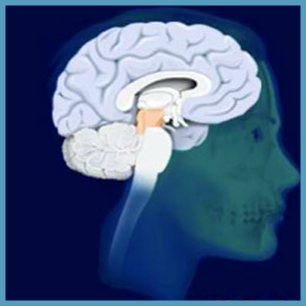
MRI gives superb contrast between different types of tissue within the human body. MRI stands for Magnetic Resonance Imaging.
By completing this module, you will learn how magnetic resonance imaging (MRI) is used in the diagnosis of many common neurological disorders, how MRI is used in imaging breast cancer, the heart, blood vessels, bile ducts and joints, and what normal and abnormal scans look like and the causes of abnormalities.
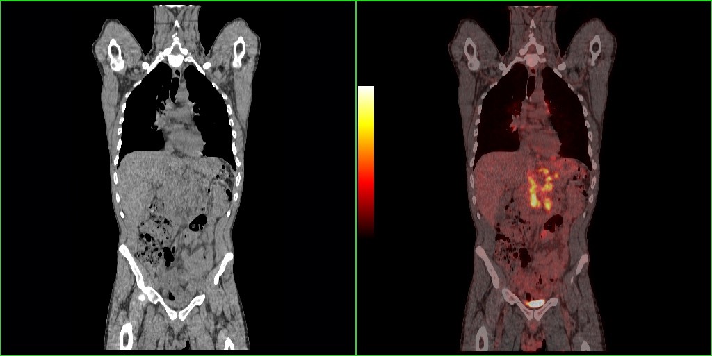
PET and SPECT imaging utilise radioactive formulations called radiopharmaceuticals in order to provide information on biological processes in the living human body.
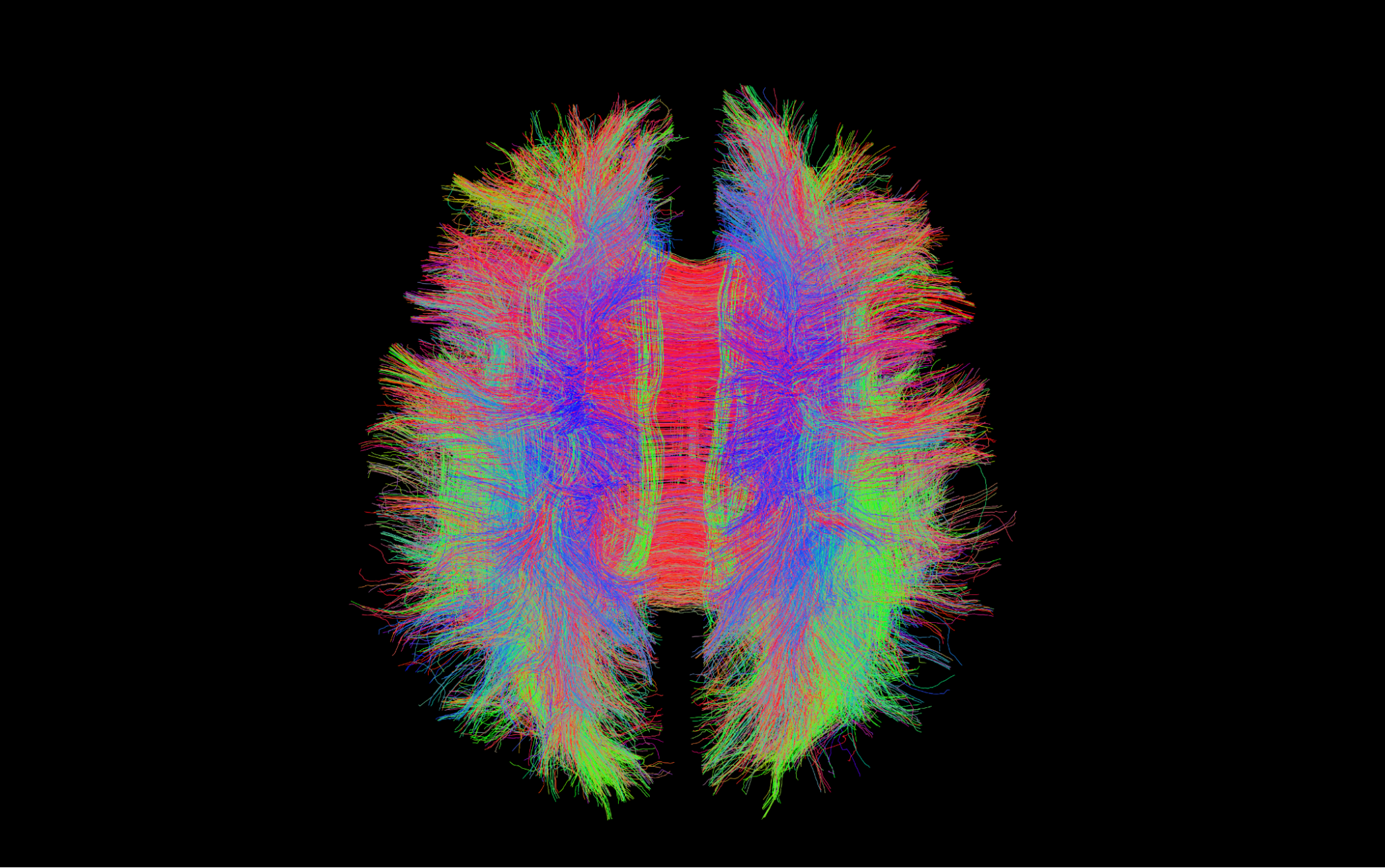
By completing this module you will learn about how MRI techniques are developing in research and in clinical practice, how MRI can be used to study blood vessels, how MRI can identify the regions of the brain that are involved in specific tasks.
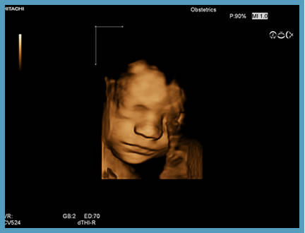
By completing this module, you will learn what ultrasound is, how and why we use ultrasound to image the body, what information ultrasound gives us about what is happening within the body, and the safety considerations when using ultrasound.
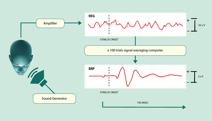
The objectives of this resource are to explain how electrical signals are generated by the brain and body, to illustrate how electrical activity in the brain and body is recorded and measured, to explain what brain functions these signals represent, and to describe examples of clinical applications.
By completing this module, you will learn about The history of CT, The nature of X-rays, The practicalities of CT, and Clinical applications of CT.
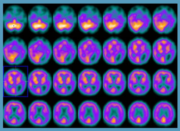
Positron emission tomography (PET) and single photon emission computed tomography (SPECT), also known as molecular imaging techniques, have in common the formation of cross-sectional images following injection of a radioactive substance. This module will illustrate the use of PET in detecting and staging cancer and the role of SPECT in the diagnosis of some common neurological disorders.

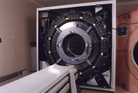
By completing this module, you will learn about Radioactivity - Atoms, Isotopes and Decay; Sources of Radioactivity; Detection of Gamma Rays; SPECT Camera Design; PET Camera Design, and Combining Function and Anatomy.
MRI is often referred to as totally safe because it does not use ionizing radiation such as X-rays. However in practice, if the correct procedures are not observed it can be extremely dangerous. As well as fatalities, burns and many other injuries have occurred.